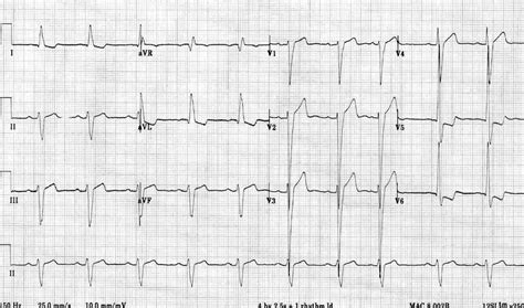
lv ecg | ecg showing lvh lv ecg The left ventricle hypertrophies in response to pressure overload secondary to conditions such as aortic stenosis and hypertension. This results in increased R wave amplitude in the left-sided ECG leads (I, aVL and V4-6) and increased S wave depth in the right-sided . Free prices and trends for Glaceon LvX Pokemon cards of the set Majestic Dawn. Free Pokemon card price guide and trends, updated hourly.
0 · signs of lvh on ecg
1 · lvh ecg pattern
2 · lvh ecg chart
3 · left ventricular hypertrophy life in the fast lane
4 · ecg showing lvh
5 · ecg lvh meaning
6 · ecg findings in lvh
7 · diagnosis of lvh on ecg
GoMobile Tires Las Vegas. 4.6 (7 reviews) Tires. “profession and a great help. I highly recommend GoMobile Tires for you tire needs, especially if you” more. Genuine Auto Services. Auto Repair. Oil Change Stations. Tires. Verified License. Online appointments. At-home services. Responds in about 2 hours. 22 locals recently requested a quote.
The left ventricle hypertrophies in response to pressure overload secondary to conditions such as aortic stenosis and hypertension. This results in increased R wave amplitude in the left-sided ECG leads (I, aVL and V4-6) and increased S wave depth in the right-sided .R Wave Peak Time Rwpt - Left Ventricular Hypertrophy (LVH) • LITFL • ECG .
ECG Pearl. There are no universally accepted criteria for diagnosing RVH in .
dior flip wallet
ECG Criteria for Left Atrial Enlargement. LAE produces a broad, bifid P wave in .In LBBB, conduction delay means that impulses travel first via the right bundle .U Waves - Left Ventricular Hypertrophy (LVH) • LITFL • ECG Library DiagnosisLeft Axis Deviation - Left Ventricular Hypertrophy (LVH) • LITFL • ECG .
The most common causes of left ventricular hypertrophy are aortic stenosis, aortic regurgitation, hypertension, cardiomyopathy and coarctation of the aorta. There are several ECG indexes, .
signs of lvh on ecg
ECG features suggestive of LV dilation (dilated cardiomyopathy) LVH is a bit of a misnomer because this actually reflects increased left ventricular mass (due to increased wall . The left ventricle hypertrophies in response to pressure overload secondary to conditions such as aortic stenosis and hypertension. This results in increased R wave amplitude in the left-sided ECG leads (I, aVL and V4-6) and increased S .

The most common causes of left ventricular hypertrophy are aortic stenosis, aortic regurgitation, hypertension, cardiomyopathy and coarctation of the aorta. There are several ECG indexes, which generally have high diagnostic specificity but low sensitivity. ECG features suggestive of LV dilation (dilated cardiomyopathy) LVH is a bit of a misnomer because this actually reflects increased left ventricular mass (due to increased wall thickness or chamber dilation). The following features may suggest LV chamber dilation: Reduction in R-wave of left precordial leads.
Left ventricular hypertrophy, especially in patients with hypertension, increases the risk of heart failure, ischemic heart disease, sudden death, atrial fibrillation and stroke.
Left ventricular hypertrophy (LVH) refers to an increase in the size of myocardial fibers in the main cardiac pumping chamber. Such hypertrophy is usually the response to a chronic pressure or volume load. The two most common pressure overload states are systemic hypertension and aortic stenosis. According to the American Society of Echocardiography and/European Association of Cardiovascular Imaging, LVH is defined as an increased left ventricular mass index (LVMI) to greater than 95 g/m in women and increased LVMI to greater than 115 g/m in men.
Left ventricular hypertrophy can be diagnosed on ECG with good specificity. When the myocardium is hypertrophied, there is a larger mass of myocardium for electrical activation to pass.
The traditional approach to the ECG diagnosis of left ventricular hypertrophy (LVH) is focused on the best estimation of left ventricular mass (LVM) i.e. finding ECG criteria that agree with LVM as detected by imaging. The principal ECG‐derived diagnostic characteristics for LVH detection include an increased QRS complex amplitude, an increased QRS complex duration, a leftward shift of electrical axis of the QRS in frontal plane, and ST‐segment deviation and T wave changes associated with left ventricular strain. 9 Furthermore, more advanced ECG methods . The ECG changes in a patient with left ventricular hypertrophy (LVH) were described 117 years ago by Einthoven in 1906 (Einthoven, 1957). He drew attention to the distinctive finding—the increased QRS amplitude in the “left hand to left foot lead” (i.e., lead III).
The left ventricle hypertrophies in response to pressure overload secondary to conditions such as aortic stenosis and hypertension. This results in increased R wave amplitude in the left-sided ECG leads (I, aVL and V4-6) and increased S .The most common causes of left ventricular hypertrophy are aortic stenosis, aortic regurgitation, hypertension, cardiomyopathy and coarctation of the aorta. There are several ECG indexes, which generally have high diagnostic specificity but low sensitivity.
dior earrings india
ECG features suggestive of LV dilation (dilated cardiomyopathy) LVH is a bit of a misnomer because this actually reflects increased left ventricular mass (due to increased wall thickness or chamber dilation). The following features may suggest LV chamber dilation: Reduction in R-wave of left precordial leads. Left ventricular hypertrophy, especially in patients with hypertension, increases the risk of heart failure, ischemic heart disease, sudden death, atrial fibrillation and stroke. Left ventricular hypertrophy (LVH) refers to an increase in the size of myocardial fibers in the main cardiac pumping chamber. Such hypertrophy is usually the response to a chronic pressure or volume load. The two most common pressure overload states are systemic hypertension and aortic stenosis. According to the American Society of Echocardiography and/European Association of Cardiovascular Imaging, LVH is defined as an increased left ventricular mass index (LVMI) to greater than 95 g/m in women and increased LVMI to greater than 115 g/m in men.

Left ventricular hypertrophy can be diagnosed on ECG with good specificity. When the myocardium is hypertrophied, there is a larger mass of myocardium for electrical activation to pass.The traditional approach to the ECG diagnosis of left ventricular hypertrophy (LVH) is focused on the best estimation of left ventricular mass (LVM) i.e. finding ECG criteria that agree with LVM as detected by imaging.
lvh ecg pattern
lvh ecg chart
The principal ECG‐derived diagnostic characteristics for LVH detection include an increased QRS complex amplitude, an increased QRS complex duration, a leftward shift of electrical axis of the QRS in frontal plane, and ST‐segment deviation and T wave changes associated with left ventricular strain. 9 Furthermore, more advanced ECG methods .

dior cruise fashion show
left ventricular hypertrophy life in the fast lane
1 . Hash House A Go Go. 4.0 (8.7k reviews) New American. Breakfast & Brunch. $$The Strip. “That said, the pancake itself was the PERFECT texture --- springy without being rubbery.moist.” more. Takeout. Start Order. 2 . Tableau. 4.2 (805 reviews) Breakfast & Brunch. New American. Bars. $$$The Strip.
lv ecg|ecg showing lvh


