lv walls | normal Lv wall thickness lv walls Segments of the left ventricle. Based on anatomical landmarks and autopsy studies (Edwards et al), the left ventricle is divided into three equal parts along . r/FinalFantasyVII. • 3 yr. ago. [deleted] 9999 damage by Tifa lv 38. Power Soul multipliers + Death Blow. Championship Belt + Gigas Armlet. FF7 ORIGINAL. Death Blow x2. Gigas Armlet +30 Str. Championship Belt +30 Str. Power Soul, +Death Sentance x2, +Low Health x4. ***Damage x16 + 60 Str. base damage like 500. Archived post.
0 · reasons for left ventricular hypertrophy
1 · normal Lv wall thickness
2 · myocardial wall
3 · lvh with repolarization abnormalities
4 · increased Lv wall thickness
5 · Lv wall thickness on echo
6 · Lv wall thickness normal values
7 · Lv wall motion abnormalities
Address: Spaces Business Centre, Hofplein 20, Rotterdam, 3032 AC, Netherlands. RegNr: 24277249. Visit: https://www.nic.lv/whois/contact/filebase.lv to contact. [Nservers] Nserver: ns1.parkingcrew.net. Nserver: ns2.parkingcrew.net. [Whois] Updated: 2024-04-12T17:12:27.431748+00:00. [Disclaimer]
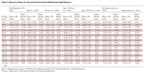
Segments of the left ventricle. Based on anatomical landmarks and autopsy studies (Edwards et al), the left ventricle is divided into three equal parts along . Recently, the consensus of the American Heart Association (AHA) 21 divided the LV into 4 walls: septal, anterior, lateral, and inferior; in turn, the .Segments of the left ventricle. Based on anatomical landmarks and autopsy studies (Edwards et al), the left ventricle is divided into three equal parts along the long axis of the ventricle. This creates three circular sections of the left ventricle named basal, mid-cavity, and apical. Recently, the consensus of the American Heart Association (AHA) 21 divided the LV into 4 walls: septal, anterior, lateral, and inferior; in turn, the 4 walls were divided into 17 segments: 6 basal, 6 mid, 4 apical, and 1 segment being the apex (Figure 2).
Left ventricular hypertrophy is a thickening of the wall of the heart's main pumping chamber, called the left ventricle. This thickening may increase pressure within the heart. The condition can make it harder for the heart to pump blood. The most common cause is .Assessment of LV function remains the most common reason for cardiac imaging because of its powerful ability to predict morbidity and mortality. Current routine methods of quantifying LV function (with LVEF) is not without limitations.Electronic calipers should be positioned on the interface between myocardial wall and cavity, and the interface between wall and pericardium. Perform at end-diastole (previously defined) perpendicular to the long axis of the LV, at or immediately below the level of . Each echocardiogram includes an evaluation of the LV dimensions, wall thicknesses and function. Good measurements are essential and may have implications for therapy. The LV dimensions must be measured when the end-diastolic and end-systolic valves (MV and AoV) are closed in the parasternal long axis (PLAX) view.

The first and most commonly used echocardiography method of LVM estimation is the linear method, which uses end-diastolic linear measurements of the interventricular septum (IVSd), LV inferolateral wall thickness, and LV internal diameter derived from 2D-guided M-mode or direct 2D echocardiography. This method utilizes the Devereux and Reichek .Each of the following echo parameters are discussed and updated in turn: left ventricular linear dimensions and LV mass; left ventricular volumes; left ventricular ejection fraction; left atrial size; right heart parameters; aortic dimensions; and tissue Doppler imaging.Wall motion is assessed in each segment of the left ventricle (Figure 1; refer to Segments of the Left Ventricle). Regional wall motion abnormalities are defined as regional abnormalities in contractile function. Ischemic heart disease is the most common cause of .The volume-based measurement of left ventricular ejection fraction (LVEF) is fundamentally different from direct measurement of myocardial motion by tissue Doppler imaging and myocardial deformation, and the reliability and precision of these measurements are also different.
Segments of the left ventricle. Based on anatomical landmarks and autopsy studies (Edwards et al), the left ventricle is divided into three equal parts along the long axis of the ventricle. This creates three circular sections of the left ventricle named basal, mid-cavity, and apical. Recently, the consensus of the American Heart Association (AHA) 21 divided the LV into 4 walls: septal, anterior, lateral, and inferior; in turn, the 4 walls were divided into 17 segments: 6 basal, 6 mid, 4 apical, and 1 segment being the apex (Figure 2).
Left ventricular hypertrophy is a thickening of the wall of the heart's main pumping chamber, called the left ventricle. This thickening may increase pressure within the heart. The condition can make it harder for the heart to pump blood. The most common cause is .Assessment of LV function remains the most common reason for cardiac imaging because of its powerful ability to predict morbidity and mortality. Current routine methods of quantifying LV function (with LVEF) is not without limitations.Electronic calipers should be positioned on the interface between myocardial wall and cavity, and the interface between wall and pericardium. Perform at end-diastole (previously defined) perpendicular to the long axis of the LV, at or immediately below the level of . Each echocardiogram includes an evaluation of the LV dimensions, wall thicknesses and function. Good measurements are essential and may have implications for therapy. The LV dimensions must be measured when the end-diastolic and end-systolic valves (MV and AoV) are closed in the parasternal long axis (PLAX) view.
The first and most commonly used echocardiography method of LVM estimation is the linear method, which uses end-diastolic linear measurements of the interventricular septum (IVSd), LV inferolateral wall thickness, and LV internal diameter derived from 2D-guided M-mode or direct 2D echocardiography. This method utilizes the Devereux and Reichek .Each of the following echo parameters are discussed and updated in turn: left ventricular linear dimensions and LV mass; left ventricular volumes; left ventricular ejection fraction; left atrial size; right heart parameters; aortic dimensions; and tissue Doppler imaging.Wall motion is assessed in each segment of the left ventricle (Figure 1; refer to Segments of the Left Ventricle). Regional wall motion abnormalities are defined as regional abnormalities in contractile function. Ischemic heart disease is the most common cause of .
gucci and louis vuitton face mask
reasons for left ventricular hypertrophy
normal Lv wall thickness
myocardial wall

On this page, you will learn how to optimise your opener and rotation in both single-target and multi-target situations. We also cover the use of your cooldowns, to ensure you can achieve the best use of them every time as a Monk DPS in Final Fantasy XIV: Endwalker (Patch 6.55).
lv walls|normal Lv wall thickness


